
Revolutionary Holotomographic 3D holographic microscope opens new era for Label-Free Live Cell Imaging
Cellular analysis plays a crucial role in a wide variety of research and diagnostic activities in the life science. However, the information available to researchers and clinicians is limited by current microscopy techniques. An innovative new tool – Holotomographic microscopy – can overcome many of these limitations and open new vistas for researchers and clinicians to understand, diagnose and treat human diseases.
Holotomographic Microscopy – New era of microscopy Tomocube's holotomography series utilize Optical Diffraction Tomography (ODT), which enables users to quantitatively and noninvasively investigate biological cells and thin tissues. ODT reconstructs the 3D refractive index (RI) distributions of live cells and by doing so, provides structural and chemical information about the cell including dry mass, morphology, and dynamics of the cellular membrane.
Videos
Files
| Attachment | Size |
|---|---|
| 18.75 MB |
News, events, promotions, webinars

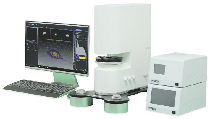
TomoStudio™, HT-1 operating SW, controls the system and visualizes the captured image in various ways. This flexible user interface provides fast imaging capability and 2D/3D/4D visualization of the cellular image based on 3D RI distributions of the cells and tissues.
TomoStudio™ works conjunctively with HT models, to visualize and analyze RI tomograms. With TomoStudio™, an user can color code the image according to refractive index and identify various types of quantitative data.
TomoStudio™ provides
- Work flow interface - User interface allows uninterrupted workflow from using the microscope to analyzing the data
- Data backup - Raw data can be stored in the computer for further analysis
- Fast image acquisition - HT-1 captures Holotomographic images every 0.4second (2.5 f/s) and 2D holographic images every 0.007second (150 f/s)
- Data analysis - Data can be processed quantitatively and real-time. User can also perform various quantitative imaging analysis
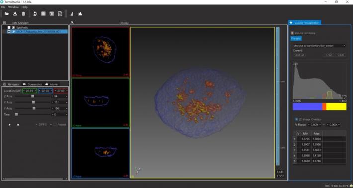
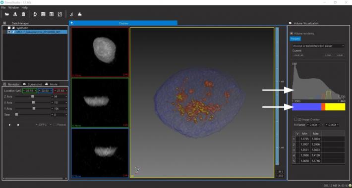
- Holographic staining - Digital color coding controller (Transfer function) is a graphical user interface to stain the sample digitally based on refractive index information.
- Dynamic image processing - Data processing does not interfere with the image acquisition process. Selective data processing is possible any time.
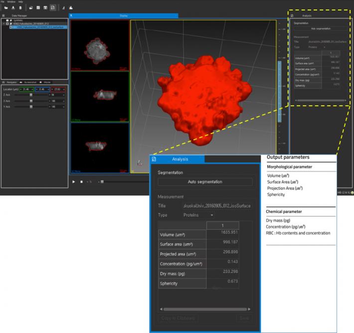
Output parameters
- Morphological parameter
- Volume (㎛³)
- Surface Area (㎛²)
- Projection Area (㎛²)
- Sphericity
- Chemical parameter
- Dry mass (pg)
- Concentration (pg/㎛³)
- RBC (Red Blood Cell): Hb contents and concentration
- Mechanical parameter
- Cell stiffness
- Dynamic membrane fluctuation
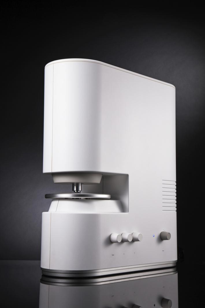
HT-1 uses holotomography technology which measures the 3D and 4D refractive index (RI) tomograms. This new technology enables quantitative, label-free measurements of live, unstained cells and tissue samples. With the dynamic micromirror device (DMD), we have eliminated moving parts from light path while image-capturing and has added unprecedented stability and precision on the outstanding performance of our product.
Simple theory
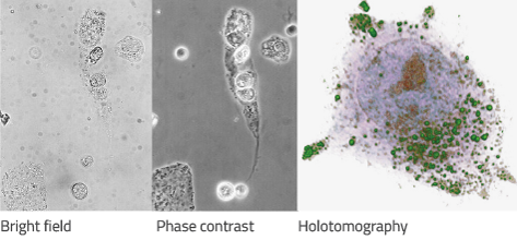
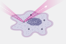
When light passes through the object, the diffraction happens according to the object's own RI(refractive index) and some properties of light including wavelength and phase shift, etc. also change. If the light that passes through the object is mixed up with the original light(reference), we can observe brightness changes in images according to the change of phase shift. This is a fundamental theory of phase contrast microscopy. Phase contrast microscopy is usually used for observation of the transparent objects (ex. Unstained biological sample, especially live cell.) which have difficulties to see without any stain or labeling.
ODT(optical diffraction tomography) is the technology that makes a 3D image (tomogram) with the RI and the 3-dimensional location which can be obtained by calculating phase shift in hologram taken around the specimen 360°. The similar technology is CT(computed tomography) using X-ray and MRI(magnetic resonance imaging) using nuclear magnetic moment.
Schematic Light path A sample is located on a stage between an objective and a condenser lens. A laser is split into a specimen and reference arm. The sample and the reference arms then generate a 2-D hologram, which is recorded by a digital image sensor. The laser illuminates the sample with the incident angle of 53°(HT-1S) or 63°(HT-1H), which rotates 360° with respect to the optical axis. A 3-D refractive index (RI) tomogram of the sample is then reconstructed from the measured multiple holograms with various illumination angles.
DMD technology
In a DMD, individual mirrors can be tilted to the angle of ±10-12°, which results to an on or off state of the reflected light. HT series utilizes a DMD in order to systematically control the illumination angle of a laser beam impinging onto a sample. Without any mechanically moving parts, the use of a DMD enables highly stable, fast, and reliable control of beam paths.
|
3-D Refractive Index: |
No labeling |
High Resolution |
|
Low Laser |
Fast in 2D/3D |
High impact, Low cost |
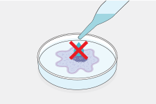
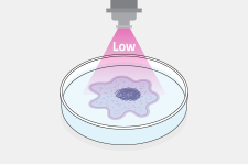

- Zero stress
Label-free imaging - Intact live cell imaging
Long-term image with short time interval - Time saving
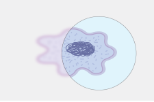
No sample preparation and Rapid 3D cell imaging - High quality 3D
Optical resolution below 200 nm (Max. 110 nm) - Quantitative bioimaging
RI enables quantitative bioimaging
(local cytoplasmic concentration, dry mass)
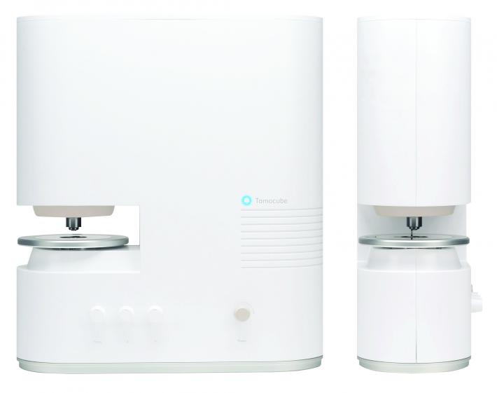
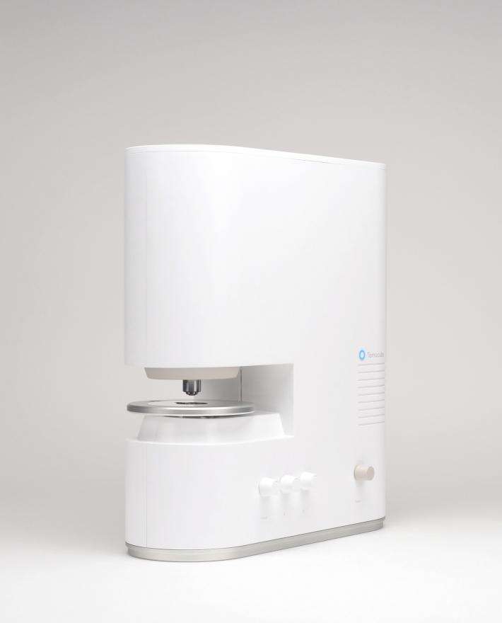
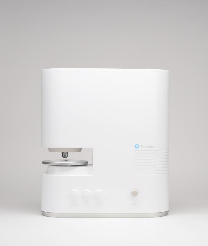
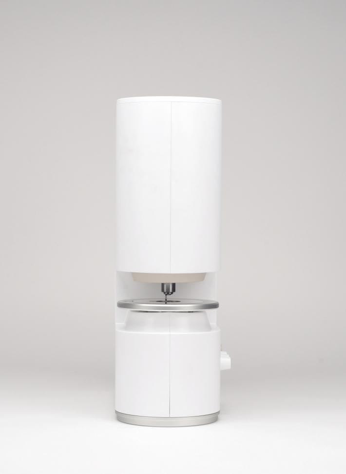
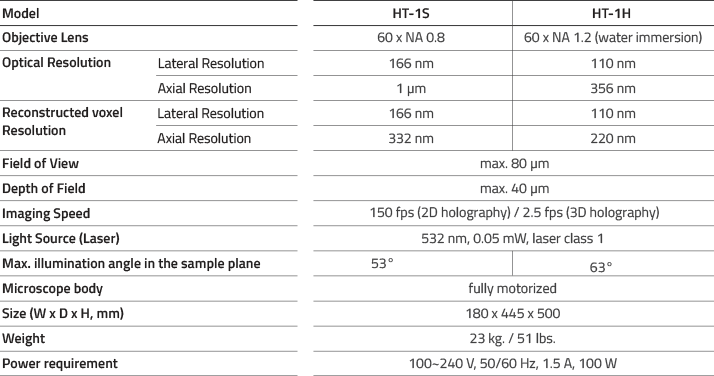
Technical Features
Environmental requirements
![]()