Brand:


Technical Features
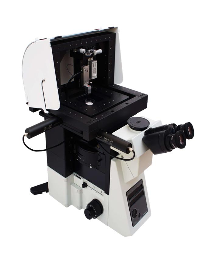
Z Stage
- Applications: Fast Z-positioning of the nanopipette; manual or motorized XY positioning (optional).
- Travel Range: 13 mm.
- Typical Step Size: 20 nm.
- Maximum Speed: 3.6 mm/min.
- Integrated Sensor: Capacitive.
- Sensor Travel Range: 25 μm.
- Positioning Accuracy: 0.1 nm.
- Linearity (closed-loop): 0.03%.
- Resonant Frequency (unloaded): 3.7 kHz.
- Resonant Frequency (200 g load): 1.7 kHz.
- Operating Temperature Range: -20°C to 80°C.
XY Stage
- Active Axes: X and Y.
- Integrated Sensor: Capacitive.
- Open-Loop Travel Range: 60×60 μm.
- Closed-Loop Travel Range: 45×45 μm.
- Resolution (Open/Closed-Loop): 0.1/0.3 nm.
- Linearity: 0.03%.
- Stiffness in Motion Direction: 10 N/μm.
- Resonant Frequency (unloaded): 1550 Hz.
- Electrical Capacitance per Axis: 9 μF.
- Dynamic Operating Current Coefficient per Axis: 25 μA/(Hz•μm).
- Operating Temperature Range: -20°C to 80°C.
- Compatibility with Optical Microscopes: Nikon Ti-U.
- Sample Dimensions: Height – 10 mm; Diameter – 35 mm; Weight – <200 g.
Mechanical Stand
- Compatibility: Integration with Nikon Ti-U, XY Stage, and Z Stage.
- Material: Non-conductive anodized duralumin.
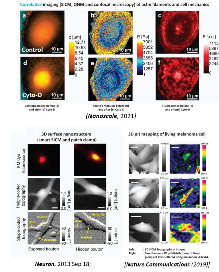
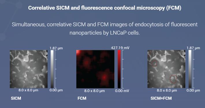
- Applications: Electromagnetic shielding; confocal microscopy; SICM; SECM; vertical patch-clamp; microinjection; localized drug delivery.
Controller
- Analog Inputs: 8 channels; 16-bit resolution; sampling rate up to 750 kHz simultaneously; voltage range from -10 to 10 V.
- Applications: Nanopipette positioning and localized delivery; electrophysiology; SPM applications; electrochemistry; sensor applications.