Enhanced Darkfield Microscope Optics

Observe nanoparticles as small as 10nm-20nm directly from your laboratory benchtop

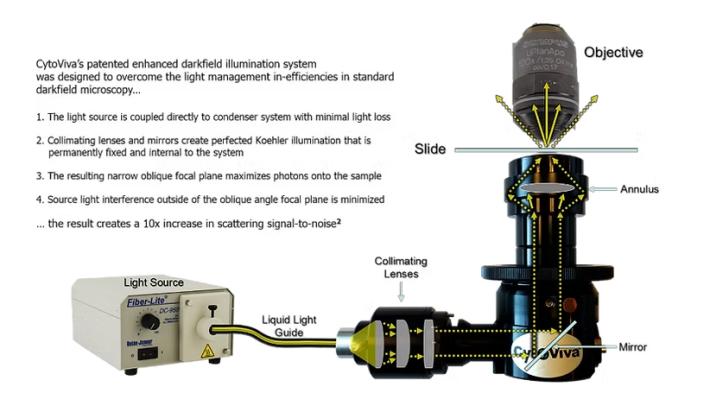
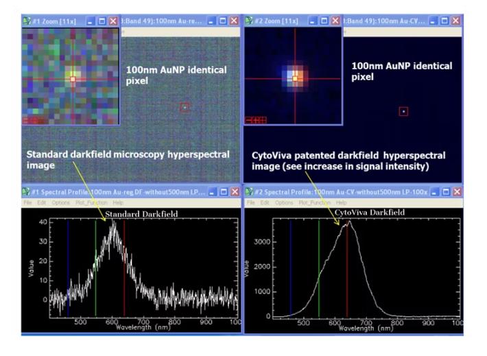
CytoViva’s darkfield microscope optics improve the signal-to-noise ratio up to ten times (10x) compared to standard darkfield optics*. This allows for imaging nanomaterials as small as 10nm-20nm directly from your laboratory benchtop**.

The CytoViva patented (U.S. patents No. 7,542,203, 7,564,623) "enhanced darkfield illumination system", which replaces the standard microscope condenser, works by coupling the light source illumination directly to the condenser optics. In this optical path, aligning and collimating lenses and mirrors fix the geometry of the light to match the condenser ring geometry. This creates a very narrow and oblique light source angle that can be precisely focused onto the sample.

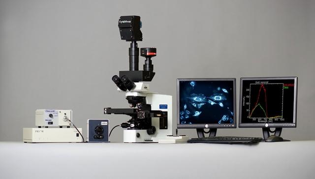
CytoViva’s darkfield microscopy enables researchers to optically observe a wide range of nanoscale materials quickly and easily; the sample can be in solution, live cell matrices, tissue, and materials. Additionally, non-fluorescent live cells and pathogens can be easily observed with a level of detail not possible with traditional optical imaging techniques such as phase contrast or differential interference contrast.

Finally, when combined with CytoViva’s hyperspectral microscopy capabilities, this high signal-to-noise ratio microscopy method allows researchers to characterize and spectrally map nanoscale samples in a wide range of environments.

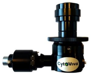
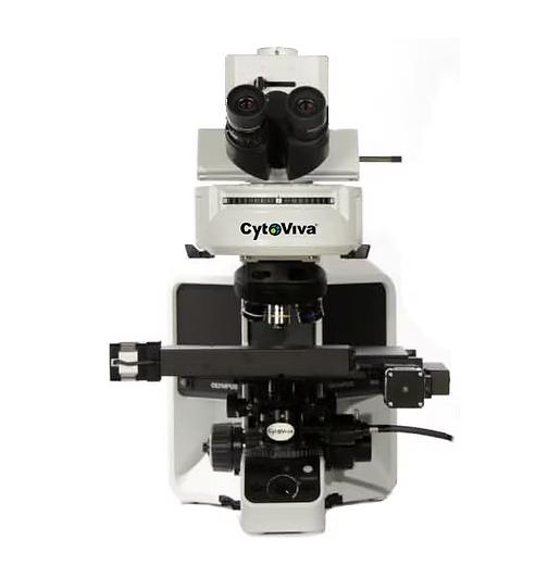
The high-resolution CytoViva illuminator is designed to fit the condenser mount of most common upright microscopes. It is the only CytoViva component that directly attaches to the microscope. Additionally, the system can be adapted to most inverted microscopes.

Zhang, P., Park, S. and Kang, S. H. (2015), Microchip Electrophoresis with Enhanced Dark-Field Illumination Detection for Fast Separation of Native Single Super-Paramagnetic Nanoparticles. Bull. Korean Chem. Soc., 36: 1172–1177.

Chaudhari, K., & Pradeep, T. (2014). Spatiotemporal mapping of three dimensional rotational dynamics of single ultrasmall gold nanorods. Scientific Reports, 4, 5948. http://doi.org/10.1038/srep05948

Videos

CytoViva’s enhanced darkfield microscope optics improve signal-to-noise up to ten times (10x) over standard darkfield optics. This enables nanomaterials as small as 10nm-20nm to be imaged right from your laboratory benchtop.
Dual Mode Fluorescence Module

Technical Features

SYSTEM PERFORMANCE
Detection of noble metals and metal oxide NPs: ~10 nm
Detection of SoftNPs (liposomes, polymers, etc.): ~75 nm

ILLUMINATION SYSTEM
Focal distance: ~1.5 mm
Field of view: ~350 μm - 400 μm
Numerical aperture: 1.2–1.4 N.A.

LIGHT SOURCE
Standard light sources: Solarc, Halogen, Mercury vapor
Power supply: 100-240Vac, 50/60 Hz, 1.6A max
Light guide type: Liquid
Outer diameter of LLG: 5 mm
Inner diameter of LLG: 3 mm

DIMENSIONS
Height (Max): 98 mm
Minimum mounting clearance: 51 mm
Dovetail angle: 30°
Minimum dovetail diameter: 41 mm
Barrel length: 110 mm
Weight: 0.43 kg

REQUIREMENTS
Immersion oil: Type A (nd > 1.515), Cargille brand
Sample prep: Cover slip - 0.17 mm thickness
Sample prep: Glass slide - 1 x 3 mm

MICROSCOPE COMPATIBILITY
Upright research microscopes
C- and O-shaped, 3-point condenser mounts
(Diameter and clearance requirements for the CytoViva optical illumination system)
Inverted research microscopes: Requires optional inverted adapter

OBJECTIVE COMPATIBILITY
Mid-range objective 10X Air
Mid-range objective 20X Air
Mid-range objective 40X Air or Water (N.A. = 0.7, Iris optional)
Mid-range objective 40X Long working distance
High-range objective 60X Oil with Iris (N.A. = ~1.4)
High-range objective 100X Oil with Iris (N.A. = ~1.4)