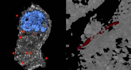
The CytoViva 3D Enhanced Darkfield Imaging System provides a method for detecting unlabeled nanostructures (particles, nanotubes, etc.) in a variety of translucent matrices (cells, tissues, organisms). This technique leverages the high signal-to-noise optical performance of the patented CytoViva Enhanced Darkfield Microscope Technology, combining it with deconvolution algorithms and particle localization methods. This allows researchers to obtain a three-dimensional optical model of their sample. Furthermore, this technique does not require fluorescent labels to visualize nanoparticles, eliminating the potential negative effects of such labels on the sample.
The 3D image is created by acquiring and storing a series of equally spaced two-dimensional images along the Z-axis. These images are then processed using CytoViva 3D Image Analysis plug-ins for ImageJ to locate particles and deconvolve surrounding cells, tissues, or other translucent matrices. The resulting optical image is then displayed in an ImageJ 3D viewer, with the possibility of further data processing in a familiar environment for many users.
The CytoViva 3D Enhanced Darkfield Imaging System is designed to facilitate research in various fields, including nanobiology (targeted drug delivery and pathogen detection) and nanotoxicology (study of nanoparticles and carbon nanotubes in tissues). Additionally, this technology is fully compatible with the CytoViva Hyperspectral Imaging System and can be used on the same microscope platform, enabling easy upgrades to existing systems to incorporate this capability.
Videos
Files
| Attachment | Size |
|---|---|
| 841.18 KB | |
| 240.58 KB |