CytoViva’s enhanced darkfield microscope optics improve signal-to-noise up to ten times (10x) over standard darkfield optics1. This enables nanomaterials as small as 10nm-20nm to be imaged right from your laboratory benchtop2.
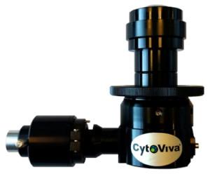
CytoViva’s patented (US patents No. 7,542,203, 7,564,623) enhanced darkfield illumination system, which replaces the standard microscope condenser, works by coupling the source illumination directly to the condenser optics. In this optical path, collimating lenses and mirrors align and fix the geometry of the light to match the geometry of the condenser annulus. This creates a very narrow, oblique angle of source illumination that can be precisely focused into the sample but bypasses the objective. The result is very intense scatter from nanoscale samples against a very dark background. Source illumination compatible with this system can be halogen, xenon or even laser, depending on the application.
CytoViva’s enhanced darkfield microscopy enables scientists to optically observe a wide range of nanoscale materials quickly and easily in solution, live cells, tissue and materials based matrices. In addition, non-fluorescent live cells and pathogens can be easily observed at a level of detail not possible with traditional optical imaging techniques such as phase contrast or differential interference contrast.
Finally, when combined with CytoViva’s Hyperspectral Imaging capability this high signal-to-noise microscopy method enables researchers to spectrally characterize and map nanoscale samples in a wide range of environments.
Videos
Files
| Attachment | Size |
|---|---|
| 647.03 KB | |
| 254.68 KB | |
| 262.3 KB |
News, events, promotions, webinars
The Dual Mode Fluorescence (DMF) module allows for the observation of both fluorescent and non-fluorescent sample portions simultaneously and in real-time. Samples are viewed directly through the microscope eyepiece and captured using a standard microscope camera without the need for complicated software or electronic manipulation.
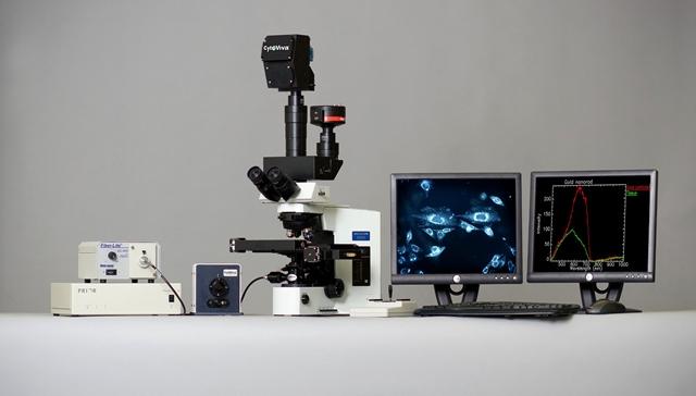
The CytoViva system is available as a comprehensive optical microscope solution. This can include an Olympus research grade microscope, camera, light source and image analysis software.
In many instances however, researchers will add the CytoViva capability to their existing microscope system. The CytoViva high resolution illuminator is designed to fit onto the condenser mount of most upright research grade microscopes. It is the only CytoViva component that attaches directly to the microscope. Additionally, the CytoViva system can be adapted for operation with most inverted microscopes.
Please contact us to determine whether your microscope is compatible with the CytoViva system.
CytoViva® provides a wide range of research grade microscope cameras available from a variety of vendors including Q Imaging and Dage MTI.
Proper camera selection will be based on your specific application and needs. For example, capturing a simultaneous, real-time image of both fluorescence and non-fluorescent sample structure will typically require a digital color camera. However, maximizing the resolution of certain images may require a cooled monochrome camera solution.
We can assist you in determining the most appropriate image capture capability for your application and provide this as part of an overall customized system solution.
To maximize the benefits of CytoViva, your microscope must be equipped with the proper objectives. Most applications will require that you have the objectives within the ranges listed below. We encourage you to contact us to determine your specific needs.
- Mid : 10x, 20x, 40x long working distance, 40x air or water NA = 0.7 iris optional
- High : 60x – 100x oil immersion NA ~1.4 w/iris
Technical Features
The patented CytoViva enhanced darkfield optical illumination system replaces the standard microscope condenser. The specialized illuminator focuses fixed-geometry, highly collimated light at oblique angles on the sample. This serves to improve signal-to-noise up to Seven (7) times over standard darkfield microscopy. This enables optimized dark field detection capability of non-fluorescing nano-scale samples.
| Noble Metals & Metal Oxide NPs | Detection ~ 10 nm |
| SoftNPs (liposomes, polymers, etc.) | Detection ~ 75 nm |
| Focal Distance | ~1.5 mm |
| Field of View | ~350 μm - 400 μm |
| Numerical Aperture | 1.2–1.4 N.A. |
| Standard Light Sources | Solarc, Halogen, Mercury Vapor |
| Power Supply | 100-240Vac,50/60 Hz, 1.6A max |
| Power | Variable Depending on Source Illumination |
| Light Guide Type | Liquid |
| Outside Diameter of LLG | 5 mm |
| Inside Diameter of LLG | 3 mm |
| Height (max) | 98 mm |
| Minimum Mounting Clearance | 51 mm |
| Dovetail Angle | 30° Slope |
| Dovetail Minimum Diameter | 41 mm |
| Barrel Length | 110 mm |
| Weight | 0.43kg (15 oz.) |
| Immersion Oil | Type A (nd> 1.515) Cargille Brand |
| Sample Prep | Cover slip - 0.17 mm Thickness |
| Sample Prep | Slides - 1 x 3 mm Glass Slide |
| Upright Research Microscopes | C-and O-shaped, 3-point Condenser Mounts (Diameter and Clearance Requirements of CytoViva Optical Illumination System) |
| Inverted Research Microscopes | Requires Optional Inverted Adapter |
| Mid Range Objective | 10X Air |
| Mid Range Objective | 20X Air |
| Mid Range Objective | 40X Air or Water (N.A.= 0.7 Iris Optional) |
| Mid Range Objective | 40X Long Working Distance |
| High Range Objective | 60X Oil with Iris (N.A.= ~1.4) |
| High Range Objective | 100X Oil with Iris (N.A.= ~1.4) |
| Warranty | One (1) year from date of purchase |
The Dual Mode Fluorescence (DMF) Module allows for the observation of both fluorescent and non-fluorescent sample portions simultaneously and in real-time. Samples are viewed directly through the microscope eyepiece and captured using a standard microscope camera without the need for complicated software or electronic manipulation.
| Technique | Transmitted Fluorescence |
| Image Modality | Fluorescence and Non-fluorescence Simultaneously in Real Time |
| Software | None Required |
| Light Source | Fluorescent Light Source |
| Lamp | Metal Arc Lamp, Mercury Vapor Lamp |
| Light Guide Type | Liquid |
| Outside Diameter | 5 mm |
| Inner Diameter | 3 mm |
| Type | CytoViva Optical Illumination System |
| Length | 129 mm |
| Width | 88 mm |
| Height | 144 mm |
| Base | 140 mm x 140 mm |
| Weight | 1.75 kg (62 oz.) |
| Excitation Filters | 4 slots |
| Excitation Filter Size | Standard 25 mm |
| Emission Filter | Triple Pass Filter |
| Emission Filter Size | Dependent on Microscope Requirements |
| Warranty | One (1) Year From Date of Purchase |