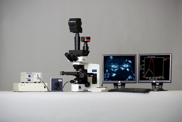
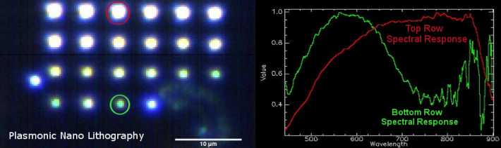
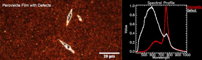
The hyperspectral microscopy technology developed by CytoViva was designed for nano-, micro-, and macro-scale samples to spectrally characterize each individual pixel of the image.
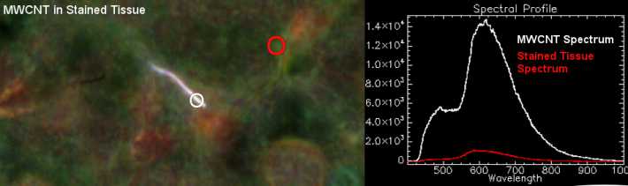
The images from the hyperspectral microscope appear very similar to those from a traditional optical microscope, but with an important difference: each pixel of a hyperspectral image provides the spectral characterization of that pixel within the VNIR or SWIR range. With 100x magnification, a hyperspectral microscope image can contain up to 700,000 pixels; from each pixel, a spectrum is generated with a 2 nm resolution, allowing for the detection of even the smallest differences in the materials that make up the sample.
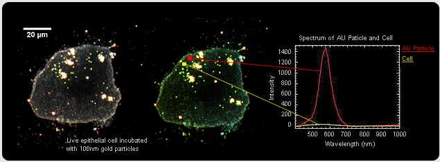
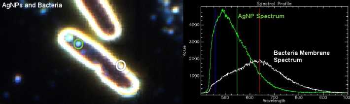
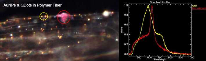
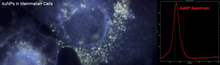
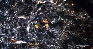
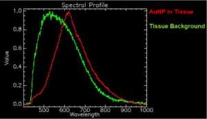
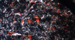
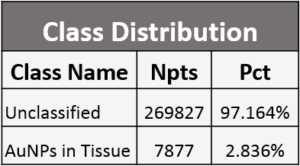
The sample can be either biological or inorganic, and may consist of simple single cells, complex matrices, or fiber layers. Among the images above, there are several examples of gold nanoparticles (AuNP) embedded in a biological sample or polymer fibers: hyperspectral imaging allows for the rapid identification and mapping of gold particles in the sample, defining both their presence and location.
These hyperspectral images are created using a linear scanning mode, moving the sample through the microscope and spectrograph's field of view via the motorized and automatic stage. The hyperspectral image is created in seconds or minutes, depending on the required exposure.
The components of the hyperspectral imaging system include a dedicated light source, an automated translational stage, a transmission diffraction grating spectrograph, and a camera. These components are integrated to interact with the acquisition software, which allows for comparing spectra within a single image or between different images. The software can also process a spectral library of materials within a sample, and using this spectral library, the same materials can be mapped in subsequent samples.
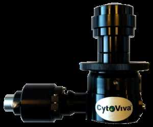
The patented dark field illumination system allows 100 times more light to be directed onto the focal plane compared to conventional dark field microscopy. As a result, the signal-to-noise ratio is the highest achievable, allowing for the detection of the finest details of the sample without the need for labeling.
Available in combination with the Enhanced Darkfield and Raman technology.
Videos
Files
| Attachment | Size |
|---|---|
| 582.29 KB | |
| 299.01 KB | |
| 595.65 KB | |
| 299.9 KB | |
| 304.41 KB | |
| 432.34 KB |
News, events, promotions, webinars
- Visualize nano-materials without using fluorescence markers or special preparation methods.
- Identify the spectrum of each individual pixel (spectral range: 400nm - 2500nm).
- Spectral libraries of pure nanoparticles are available, enabling the identification of specific materials in complex matrices and their unambiguous localization.
- An environmental control chamber is available for long-term studies on live cells.
- It is suitable for applications such as nanoparticle mapping within tissues, drug delivery, nano-toxicity assays, pathogen identification, and more.
Technical Features
- Spatial Resolution: 100 nm
- Spectral Resolution: 2 nm
- Exposure Time: 5µs – 60 sec.
- Light Source: Quartz Halogen with Aluminum Reflector
- Wavelengths:
- VNIR (400 nm – 1,000 nm)
- SWIR (900 nm – 1,700 nm)
- Maximum Spatial Scan Width: 896 µm with 10x objective
- Pixel Size: 6.45 µm x 6.45 µm
- Motorized Stage:
- Travel Range: 114 mm x 75 mm
- Motion Resolution: 10 nm
- Analysis Software: ENVI 4.8
- Spectral Image Display: Real-time display of the recreated RGB image from spectral data