

Files
| Attachment | Size |
|---|---|
| 652.91 KB |
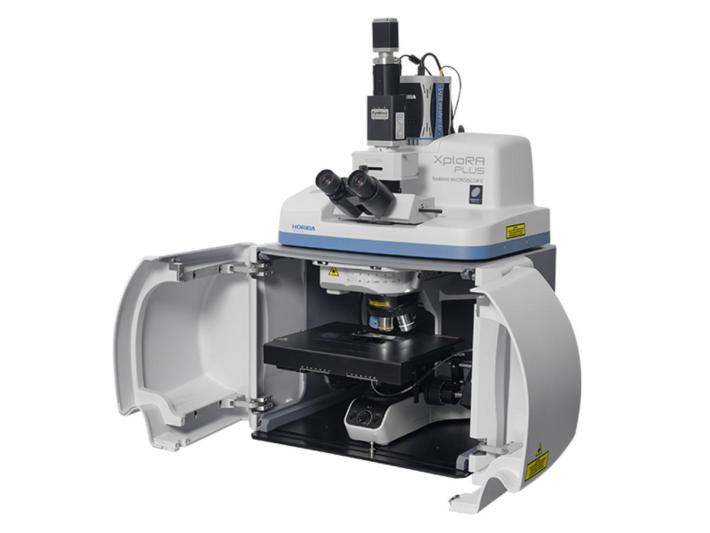
Technical Features
Raman Component Specifications
Laser Options
- Integrated up to 3 internal lasers: 532 nm, 638 nm, 785 nm
- Other wavelengths and high-power options available upon request
- PC controlled with AutoSwitch option for Raman/white light selection
Spectral Resolution: 1.4 cm⁻¹ to 8 cm⁻¹ depending on grating, laser, and CCD selection
Spectral Range: 50 cm⁻¹ to 4000 cm⁻¹ depending on grating and laser selection
Spectrograph
- Flat-field spectrometer for imaging, compatible with large CCD detectors
- High throughput with 4-position grating turret
Confocal Resolution
- Fully confocal, adjustable confocal aperture, 500 nm XY resolution
- SWIFT™ mode option for 10x faster Raman imaging
Detector
- OE. TE deep-air cooled scientific CCD (-60°C), 1024 x 256 pixels
- 16-bit, up to 1.48 MHz readout speed
Vertical Research Microscope
- Standard 2-position illuminator with transmission/reflection illumination
- USB image camera for image acquisition
- Included objectives: 10x and 100x
Optical Imaging
- Standard epi-illumination
- Advanced darkfield illumination for transmission
Objective Sampling Optics
- Long working distance options: 50x and 100x
- Macro cuvette cell sampler
- Fibre-optic probes
Software
- OneClick functionality as standard
- LabSpec Spectroscopy Suite
- Available options: database, chemometrics, imaging, ParticleFinder
Weight: 35 kg
Operating Temperature: 15 - 28°C (optimal 22°C ± 1°C)
Voltage: 110/240 VAC, mains supply
Dimensions (WxDxH): 479 x 352 x 661 mm
Note: No water cooling or LN2 supply required
Hyperspectral Component Specifications
Spatial Resolution: 100 nm
Spectral Resolution: 2 nm
Exposure Time: 5 µs – 60 sec
Light Source: Quartz halogen with aluminum reflector
Wavelengths: VNIR (400 nm – 1,000 nm)
Maximum Spatial Scan Width: 896 µm with 10x objective
Pixel Size: 6.45 µm x 6.45 µm
Motorized Stage:
- Travel Range: 114 mm x 75 mm
- Motion Resolution: 10 nm
Analysis Software: ENVI 4.8
Spectral Image Display: Real-time display of the RGB image recreated from spectral data
CytoViva Enhanced Darkfield Optical Illumination System Specifications
System Performance:
- Noble metals & metal oxide NPs detection: ~10 nm
- SoftNPs (liposomes, polymers, etc.) detection: ~75 nm
Illumination System:
- Field of View: ~400 μm
- Numerical Aperture: 1.2–1.4 N.A.
Light Source:
- Standard Light Sources: Solarc, halogen, mercury vapor
- Power Supply: 100-240V AC, 50/60 Hz, 1.6A max
- Light Guide Type: Liquid (5 mm OD, 3 mm ID)
Dimensions:
- Maximum Height: 98 mm
- Minimum Mounting Clearance: 51 mm
- Weight: 0.43 kg
Requirements:
- Immersion Oil Type A (nd > 1.515), Cargille Brand
- Cover Slip Thickness: 0.17 mm
- Glass Slide: 25 mm x 75 mm x 1 mm
Microscope Compatibility:
- Upright Research Microscopes:
- C- and O-shaped, 3-point condenser mounts
- (Diameter and clearance requirements for CytoViva optical illumination system)
- Inverted Research Microscopes: Requires optional inverted adapter
Objective Compatibility:
- Low Range Objective: 10X Air, 20X Air
- Mid Range Objective: 40X Air (N.A. = 0.7, optional iris), 40X Long Working Distance
- High Range Objective: 60X and 100X Oil with Iris (N.A. ~1.4)