Holotomography is a technique that uses low-intensity light to acquire the refractive index of cells from multiple angles. This technology has emerged as an excellent solution for cellular imaging, enabling high-resolution images while preserving cell health by using low-intensity light sources. Tomocube's second-generation holotomography, the Tomocube HT-X1, employs low-coherence light sources, ensuring lower toxicity and noise-free high-resolution image acquisition compared to laser-based methods.
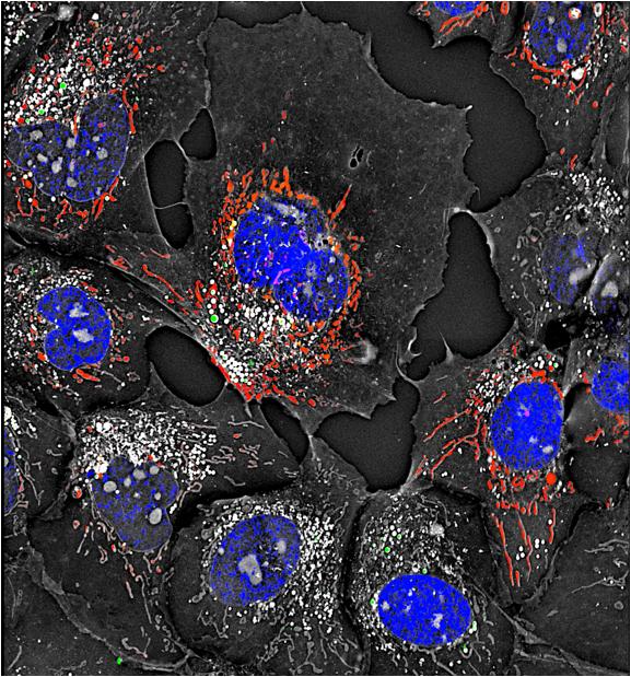
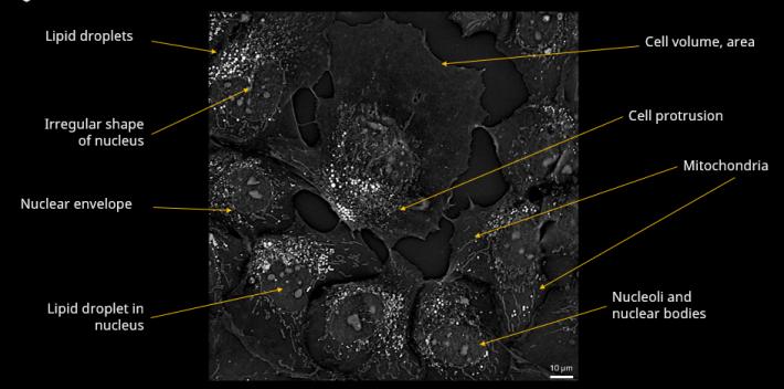
The high-resolution images obtained through this technology allow real-time observation not only of the morphology of live cells but also of the structure of subcellular organelles, such as the nucleus, nucleolus, mitochondria, and lipid particles. The integrated incubator provides a stable culture environment, enabling prolonged monitoring of sensitive cells, such as stem cells or organoids.
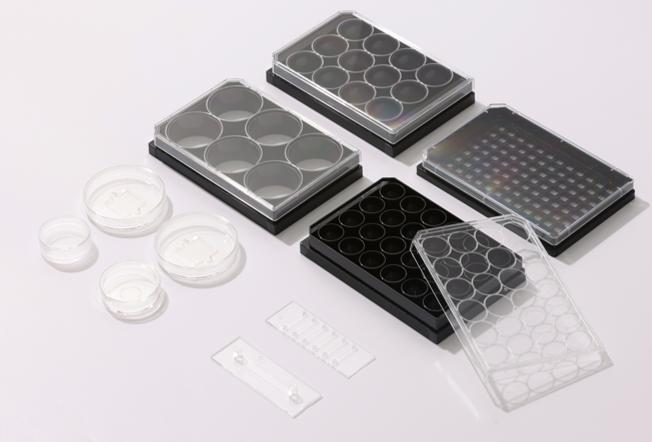
This innovative system is compatible with Tomocube’s supports as well as with various commercial imaging plates, supporting analysis up to 96-well plates, making it ideal for high-throughput screening. Using the HT-X1 allows for the acquisition of high-resolution images of cells, microorganisms, organoids, and tissue samples. Additionally, the holotomography analysis software, TomoAnalysis, allows obtaining quantitative information based on the refractive index, including high-resolution 3D images of cells, as well as data on volume, area, concentration, and dry mass. Therefore, using the HT-X1 proves to be a crucial tool for understanding biological phenomena.
The holotomography approach in live cell imaging does not require fixation or labeling, reducing unwanted artificial interventions and the need for prolonged exposure to light. By minimizing the risks of phototoxicity and photobleaching, the sample remains in its natural state for observation. With a rapid acquisition speed of 1 second per frame, HT enables real-time monitoring of dynamic cellular processes with exceptional temporal resolution.
- Label-free 3D visualization: Label-free 3D live cell imaging of monolayered cells and 3D organoids
- Correlative fluorescence: Correlative fluorescence imaging to jointly obtain biomolecular specificity information
- Long-term timelapse: Built-in incubator that provides a stable cell culture environment
- High-throughput screening: Multi-well plate compatibility for high-throughput experiments
- Quantitative analysis: Quantitative measurements analysis of cells and subcellular components
Videos
Files
| Attachment | Size |
|---|---|
| 2.45 MB | |
| 5.59 MB |
News, events, promotions, webinars

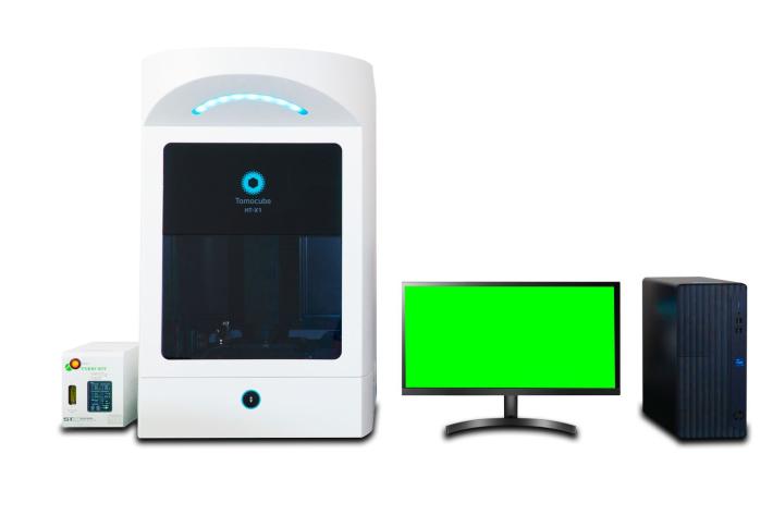
1. HT-X1 main unit
- Holotomography Illumination
- Camera
- Stage
- Incubator
- Fluorescence
2. Environmental control unit
- External Regulator for Temperature and CO2
3. Software
- TomoStudio X for acquisition control
- TomoAnalysis for data analysis and reporting


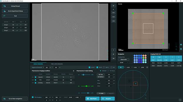
As the control software for the HT-X1, TomoStudio X is designed to operate the system as you design the experiment. TomoStudio X offers flexible workflows for multi-dimensional image acquisition of holotomography and fluorescence in multi-well imaging conditions. All the essential control and status of the system are displayed all at one glance.
Functions
- Data acquisition
- Experiment setting
- Timelapse sequence
Technical Features
HT-X1
- Objective Lens: 40x 0.95 NA air
- Camera: 2.8 Megapixel CMOS
- Compatible Supports: Dishes, Plates, Slides
- Lateral Resolution: 156 nm
- Axial Resolution: 803 nm
- Axial Range: 140 µm
- Field of View: 218 µm × 165 µm
- Imaging Modes: Holotomography, Brightfield (Grayscale), Fluorescence
- HT Light Source: LED 450 nm
- Fluorescence: Up to 4 channels – Hoechst, FITC, TRITC, Cy5
- Stage: Motorized with Laser Autofocus Module
- Minimum Acquisition Time: 3.5 seconds/image
- Dimensions: 921 × 565 × 732 mm (H, W, D)
- Weight: 90 kg
ENVIRONMENTAL CONTROLLER
- Temperature Range
- Sample Temperature: 37 °C
- Top Heater: 10 °C – 65 °C
- Bath Heater: 10 °C – 50 °C
- Stage Heater: 10 °C – 50 °C
- Lens Heater: 10 °C – 45 °C
- CO2 Range: 5-20% ±0.1%
- Dimensions: 151 x 263 x 196 mm (H, W, D)
- Weight: 3.8 kg
SYSTEM
- Operating System: Windows 10 IoT
- CPU: Intel Core i7 or equivalent
- GPU: NVIDIA GeForce RTX 4090 or equivalent
- RAM: 128 GB
- Screen: QHD (2560 × 1440)