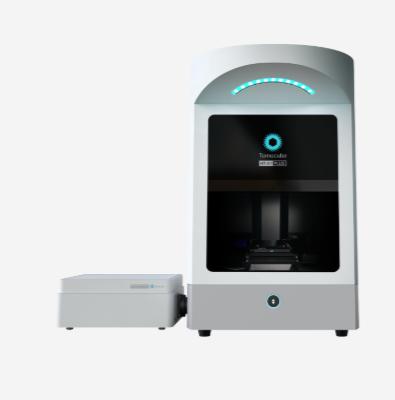
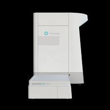
HT-X1™ Plus takes bioimaging to the next level, building on the proven success of the HT-X1™.
The HT-X1™ Plus advances high-resolution holotomography even further, offering improvements designed to meet the ever-evolving needs of biomedical research. Discover clearer, more detailed imaging of multi-layered specimens with improved illumination optics and advanced image reconstruction algorithms.
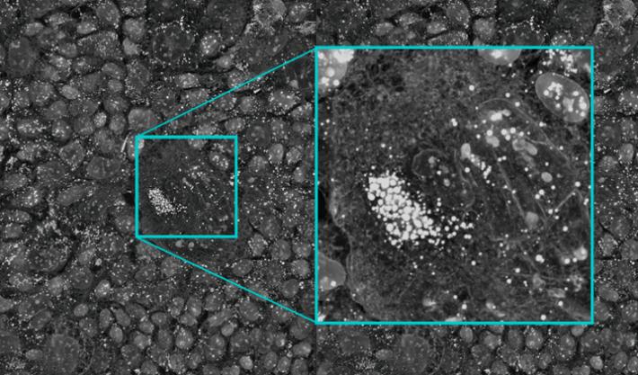
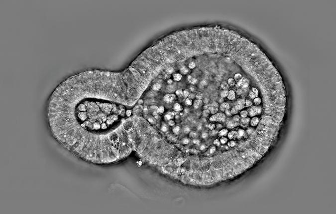
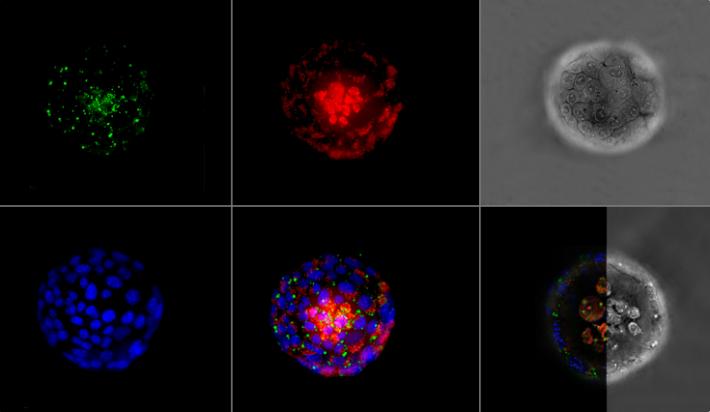
Equipped with a high-performance camera with a 4x larger field of view and significantly reduced acquisition times, the HT-X1™ Plus is perfect for high-throughput phenotypic screening of cells, dense organoids, tissue sections, and fast-moving microorganisms. The platform integrates long-wavelength light sources, improving penetration depth and reducing scattering noise to achieve clearer 3D visualization. Its enhanced correlative imaging capabilities, incorporating an sCMOS-based fluorescence module, enable seamless integration of molecular studies with high-resolution 3D images at single-cell resolution.
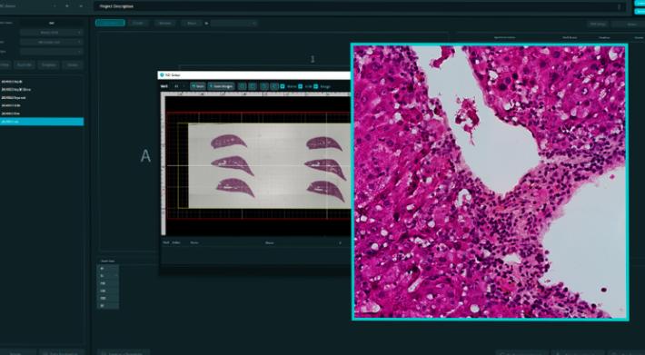
With the new color brightfield imaging modality and wide preview scan features, researchers can gain deeper insights into tissue section studies. The platform allows for the seamless integration of complex structural data, obtained through 3D optical sectioning, with histological information from H&E staining or immunohistochemistry. This integration enhances our understanding of tissue morphology and dynamics, accelerates advancements in clinical pathology and diagnostics, and helps pave the way for the future of personalized medicine.
HT-X1™ Plus is optimized for high-throughput screening, making it particularly well-suited for high-content image-based drug screening research. Featuring a high-performance CXP camera and AI-powered image reconstruction algorithms, the platform excels in both coverage and acquisition speed. Its large field of view of 308 μm x 308 μm and rapid 3D scanning capability allow researchers to efficiently analyze an entire 96-well plate in less than 30 minutes. This efficiency enables large-scale experiments with unmatched precision and consistency, leading to faster and more reliable data acquisition that significantly accelerates the drug discovery process.
Large Field of View: Capture expansive areas without the need for stitching, ideal for large-scale, high-content experiments.
Faster Image Acquisition: Efficiently acquire high-content images, making the HT-X1™ Plus perfect for high-throughput screening and dynamic specimen monitoring.
Flexible Choice of Light Source: Customize your imaging with three wavelength options to enhance contrast or improve penetration, tailored to your research needs.
Combine Advanced Fluorescence: Maximize 3D imaging quality by integrating the HT with the sCMOS-equipped fluorescence module, delivering cutting-edge 3D fluorescence imaging.
Wide Preview + Color Brightfield: Gain deeper insights into tissue section studies with the wide preview scan mode, paired with correlative color brightfield imaging.
Videos
Files
| Attachment | Size |
|---|---|
| 10.31 MB |
News, events, promotions, webinars

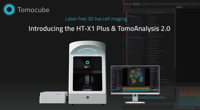
As the control software for the HT-X1™ Plus, TomoStudio X is designed to manage the system throughout the experiment setup process. TomoStudio X provides flexible workflows for acquiring multidimensional holotomography and fluorescence images. All essential controls and system status are displayed on a single screen.
Features:
- Data Acquisition
- Experiment Setup
- Timelapse Sequencing
Technical Features
HT-X1 Plus
- Objective Lens: 40x 0.95 NA air
- Camera: 20 Megapixel CMOS, CXP-12
- Supports: Dishes, Plates, Slides
- Lateral Resolution: 156 nm
- Axial Resolution: 803 nm
- Axial Range: 140 µm
- Field of View: 308 µm x 308 µm
- Imaging Modes: Holotomography, Brightfield (color), Fluorescence
- HT Light Source: LEDs 444, 520, 660 nm
- Fluorescence: Up to 4 channels – Hoechst, FITC, TRITC, Cy5
- Stage: Motorized with Laser Autofocus Module
- Minimum Acquisition Time: 0.5 seconds/image
- Dimensions: 921 x 565 x 732 mm (H, W, D)
- Weight: 95 kg
FLUORESCENCE MODULE
- FL Light Source: LEDs
- Filter Change Time: 100 ms
- Fluorescence Image Sensor: sCMOS
- Quantum Efficiency: 95% (at a wavelength of 580 nm)
- Dimensions: 174 x 434 x 174 mm (H, W, D)
- Weight: 25 kg
ENVIRONMENTAL CONTROLLER
- Temperature Range
- Sample Temperature: 37 °C
- Top Heater: 10 °C – 65 °C
- Bath Heater: 10 °C – 50 °C
- Stage Heater: 10 °C – 50 °C
- Lens Heater: 10 °C – 45 °C
- CO2 Range: 5-20% ±0.1%
- Dimensions: 151 x 263 x 196 mm (H, W, D)
- Weight: 3.8 kg
SYSTEM
- Operating System: Windows 10 IoT
- CPU: Intel Core i7 or equivalent
- GPU: NVIDIA GeForce 6000 Ada or equivalent
- RAM: 128 GB
- Screen: QHD (2560 x 1440)